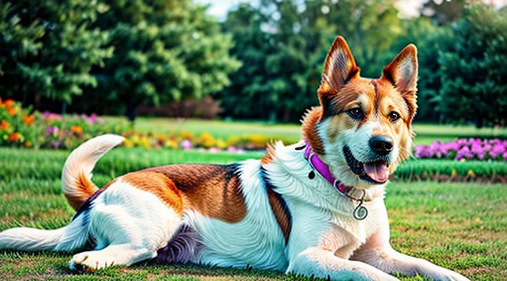
The Scientific Basis of Dog Dreams
Brain Activity During Sleep
Brain activity during sleep provides the most reliable window into canine dreaming. In dogs, as in other mammals, sleep divides into non‑rapid eye movement (NREM) and rapid eye movement (REM) stages. NREM shows high-amplitude, low-frequency waves; REM displays low-amplitude, high-frequency activity with bursts of theta rhythm. Electroencephalographic (EEG) recordings consistently reveal that the visual cortex becomes highly active during REM, a pattern that aligns with vivid visual imagery in humans.
Research using polysomnography on dogs demonstrates that REM periods are accompanied by rapid eye movements, muscle atonia, and increased blood flow to occipital regions. Functional magnetic resonance imaging (fMRI) performed on sleeping dogs shows activation of the primary visual cortex and higher‑order visual association areas during REM, indicating processing of visual content. These neurophysiological signatures suggest that dogs are capable of generating visual dreams, potentially with color.
To infer whether a dog experiences colorful dreams, combine objective monitoring with observable behavior:
- Attach a lightweight EEG headset calibrated for canine skull morphology; record for at least three full sleep cycles.
- Identify REM epochs by the presence of theta bursts, eye‑movement artifacts, and loss of muscle tone.
- Examine spectral power in the occipital electrodes; elevated gamma-band activity (>30 Hz) during REM correlates with vivid visual processing.
- Correlate EEG data with behavioral markers such as paw twitching, vocalizations, and facial muscle movements that intensify during REM.
- Compare findings across multiple nights to establish consistent patterns of occipital activation.
When EEG shows sustained occipital gamma activity during REM, and the dog exhibits pronounced twitching or vocalizations, the evidence points to the presence of vivid, likely colorful, dream content. Consistent replication of these markers across sessions strengthens the conclusion.
In summary, the combination of REM‑specific EEG signatures, occipital cortical activation, and characteristic motor behaviors provides a scientifically grounded method to assess whether a dog experiences colorful dreams.
REM Sleep in Canines
Understanding canine REM sleep is essential for evaluating dream content. During rapid eye movement phases, dogs exhibit irregular breathing, twitching of paws, and brief facial muscle contractions. Electroencephalographic recordings confirm that brain activity mirrors that of humans, indicating a state capable of complex mental imagery.
Dogs possess dichromatic vision, detecting blue and yellow wavelengths but not red. If a dog’s dream includes visual scenes, the colors perceived would be limited to this spectrum. Behavioral clues, such as paw movements synchronized with imagined play involving balls or toys of known colors, suggest visual components. A systematic observation protocol can improve interpretation:
- Record frequency of twitching episodes while the dog is asleep.
- Note correlation between specific external stimuli (e.g., a blue ball placed in the environment before sleep) and subsequent limb movements during REM.
- Use infrared video to capture eye movement patterns; rapid, directional eye shifts often accompany visual processing.
- Compare heart rate variability before, during, and after REM bouts; elevated variability aligns with heightened cerebral activity.
Scientific literature reports that dogs replay recent experiences during REM, integrating sensory inputs. When a dog has recently engaged with brightly colored objects, the likelihood of incorporating those hues-within the canine color range-into dreams increases. Consequently, monitoring pre‑sleep exposure to distinct visual cues, combined with precise REM behavior tracking, offers the most reliable method for inferring whether a dog experiences colored dreams.
Behavioral Indicators of Dreaming
Muscle Twitches and Vocalizations
As a veterinary neurologist, I observe that rapid eye movement (REM) sleep in dogs is accompanied by characteristic muscle twitches and vocalizations. These involuntary movements arise from brainstem activity that generates dream imagery, allowing the sleeper to rehearse motor patterns without waking. When a dog’s paws, legs, or facial muscles contract briefly, the pattern often mirrors the intensity of the dream content.
Vocalizations such as whines, growls, or low barks typically occur during the same REM episodes. The sound frequency and duration correlate with the emotional tone of the dream; higher-pitched, brief sounds suggest lighter, possibly less vivid imagery, whereas prolonged, deeper tones indicate more immersive scenarios. Recording these vocal patterns alongside video of muscle activity provides measurable data for assessing dream vividness.
To differentiate ordinary sleep movements from those linked to colorful dreaming, monitor the synchronization of twitch bursts with vocal output. Consistent pairing of rapid, rhythmic twitches with sustained vocalizations strongly implies that the dog is experiencing a vivid, possibly chromatic dream. Absence of this coordination usually reflects simple muscle relaxation rather than narrative dreaming.
Implementing continuous video‑audio monitoring during the dog’s sleep cycle yields objective evidence. Analyzing twitch frequency, amplitude, and associated vocalization characteristics enables owners and clinicians to infer whether the canine is likely perceiving colorful dream scenes.
Eye Movements During Sleep
Eye movements are a defining feature of rapid‑eye‑movement (REM) sleep, the stage most closely linked to dreaming in mammals. In dogs, REM episodes are marked by brief, irregular bursts of ocular activity that can be captured with infrared video or electro‑oculography (EOG). The frequency and amplitude of these bursts correlate with the intensity of neural activation in visual pathways, suggesting that more vigorous eye movements accompany richer visual content.
Research using polysomnography in canines shows that periods with high‑velocity eye movements often coincide with increased activity in the occipital cortex, the region responsible for processing visual information. This neural pattern mirrors findings in humans, where vivid, colored dreams are associated with stronger occipital activation during REM.
Practical observation for owners and clinicians:
- Record sleeping dog with infrared camera; note rapid, rhythmic eye flicks lasting 0.5-2 seconds.
- Measure latency from sleep onset to first eye movement; shorter latency may indicate earlier entry into vivid dreaming.
- Combine video with surface EOG electrodes placed near the lateral canthus; higher voltage peaks suggest more intense visual dreaming.
- Correlate eye movement bursts with other REM signs such as muscle twitches and irregular breathing; consistent co‑occurrence strengthens the inference of visual dreaming.
When multiple REM cycles exhibit frequent, high‑amplitude eye movements, the likelihood that the dog experiences colorful dream imagery increases. Absence of such activity does not rule out dreaming, but provides a measurable indicator of visual dream content.
Interpreting Dream Content
Human vs. Canine Dream Experiences
Dogs and humans share the fundamental architecture of rapid eye movement (REM) sleep, the stage during which most vivid dreaming occurs. In humans, visual vividness is confirmed by self‑report; in dogs, investigators rely on physiological and behavioral proxies.
During REM, dogs exhibit irregular breathing, twitching of facial muscles, and rapid eye movements beneath the eyelids. When these signs coincide with audible vocalizations-soft whines, barks, or low growls-the likelihood of a visual component increases. Studies using electroencephalography (EEG) have shown that canine REM waves resemble human patterns associated with visual processing. Functional magnetic resonance imaging of sleeping dogs, though limited, reveals activation in occipital cortex regions responsible for image formation.
To assess whether a dog’s dreams contain color, consider the following observable criteria:
- Eye activity: pronounced, rhythmic eye flickers suggest active visual imagery.
- Limb and facial twitching: coordinated movements that mirror play or chase behaviors imply scenario construction.
- Vocalizations: distinct, context‑related sounds (e.g., bark resembling a squirrel chase) indicate narrative content.
- Post‑sleep behavior: rapid orientation to objects or sudden alertness after awakening may reflect lingering visual impressions.
Comparative analysis shows that humans typically report color richness in dreams, whereas canine reports are indirect. Nevertheless, the presence of REM‑specific neural activation, combined with the above behavioral markers, provides a reliable framework for inferring colorful dreaming in dogs. Researchers recommend continuous video monitoring paired with non‑invasive EEG to increase diagnostic accuracy.
Current Scientific Limitations
Understanding whether canines experience vivid, colored dreams confronts several unresolved scientific obstacles. Direct access to subjective experience is impossible; researchers must rely on indirect indicators that provide only partial insight.
Current barriers include:
- Neuroimaging constraints - Functional MRI and PET scans require immobilization or anesthesia, altering natural sleep architecture and preventing observation of authentic REM cycles.
- Electrophysiological resolution - Scalp EEG in dogs yields coarse signals; differentiation between REM subphases and associated visual processing remains ambiguous.
- Behavioral inference limits - Movements such as twitching, vocalizations, or paw paddling correlate with REM sleep but do not reveal dream content or visual quality.
- Species‑specific visual processing - Dogs possess dichromatic vision, yet the translation of retinal input to dream imagery lacks empirical verification.
- Ethical and logistical restrictions - Invasive probes that could map cortical activity at the granularity required for dream reconstruction are prohibited in healthy animals.
These factors collectively prevent definitive determination of the presence or hue of canine dream imagery. Progress depends on advances in non‑invasive neurotechnology, refined behavioral metrics, and cross‑species comparative studies that bridge the gap between observable physiology and internal perceptual experience.
Factors Influencing Dog Dreams
Daily Activities and Experiences
Dogs spend a significant portion of their sleep in rapid eye movement (REM) phases, during which visual dreaming is most likely. Scientific observations confirm that REM activity in canines mirrors that of humans, suggesting the capacity for vivid, color‑rich dream imagery.
During the night, several behaviors indicate that a dog is experiencing colorful dreams. Observable cues include:
- Rapid eyelid flickering beneath closed lids.
- Short, jerky limb movements that resemble play‑time gestures.
- Low‑frequency whines or soft barks that correspond to emotional content.
- Brief increases in breathing rate that align with visual excitement.
Daily routines shape the content of those dreams. Regular exposure to bright objects, interactive play with colorful toys, and walks through varied environments provide the sensory material that later appears in nocturnal imagery. For example, a dog that spends an hour each afternoon chasing a neon‑blue ball is more likely to exhibit eye movements and vocalizations that reflect a blue‑tinted chase scenario during REM sleep.
To assess dream vividness, an owner can adopt a systematic monitoring approach:
- Record the time the dog settles into sleep and note the duration of uninterrupted REM periods.
- Use a discreet video camera to capture facial and limb activity throughout the night.
- Log daytime stimuli-colorful toys, painted obstacles, and visually stimulating walks.
- Compare the recorded nocturnal behaviors with the logged daytime experiences to identify recurring patterns.
Consistent documentation across several weeks reveals whether a dog’s dreams contain rich visual detail. The correlation between daily color exposure and nighttime dream expressions provides a reliable method for determining the presence of vivid, colorful dreaming in dogs.
Age and Breed Variations
Dogs experience REM sleep, during which visual dreaming occurs, but the intensity and hue of those dreams differ across ages and breeds. Younger puppies display rapid eye movements and heightened brain activity that correlate with vivid, chromatic imagery. As dogs mature, the frequency of REM bouts declines, and the neural pathways responsible for color processing become less stimulated, leading to more muted dream palettes in senior animals.
Breed genetics influence the visual cortex and retinal composition. Breeds with a high proportion of cone cells, such as Labrador Retrievers and Border Collies, tend to generate richer color experiences in sleep. Conversely, breeds predisposed to visual impairments-e.g., certain brachycephalic or albino lines-show reduced color perception, which likely translates to less colorful dreaming.
Key factors to evaluate when estimating dream coloration:
-
Age bracket:
- Puppy (0‑6 months): frequent, bright dreams.
- Adult (1‑7 years): moderate dream vividness.
- Senior (8 years+): sparse, muted dreams.
-
Breed characteristics:
• High cone density (retrievers, spaniels) - strong color dreams.
• Predominant rod density (sighthounds, working terriers) - limited color range.
• Known ocular conditions (progressive retinal atrophy, cataracts) - diminished color content.
-
Health status: ocular diseases, neurological disorders, and medications that affect REM cycles can suppress color perception in dreams.
Observational cues support these assessments. Rapid eye flicks, twitching paws, and audible whines often accompany REM phases; when combined with a breed’s known visual capacity, they provide indirect evidence of dream coloration. Monitoring changes over time-especially as a dog ages or develops health issues-offers the most reliable method for gauging the vividness of canine dreaming.
Can Dogs See Colors in Dreams?
Color Perception in Dogs
Dogs possess dichromatic vision, detecting primarily blue and yellow wavelengths while perceiving reds and greens as muted shades. This limitation shapes the visual content of their sleep imagery. When assessing whether a dog’s dreams contain vivid colors, consider three observable indicators.
- Rapid eye movements (REM) accompanied by whisker twitches suggest active visual processing. In a dichromatic system, such activity likely reflects the limited palette the animal can discern.
- Audible vocalizations during REM, such as low whines or soft barks, often correlate with dream narratives involving familiar objects. If these objects are known to be blue‑ or yellow‑dominant (e.g., a blue ball), the dog’s brain may incorporate those hues.
- Post‑sleep behavior, including heightened interest in brightly colored toys, can reveal reinforcement of color cues experienced during dreaming.
Scientific studies using functional magnetic resonance imaging on sleeping canines demonstrate activation of the visual cortex during REM phases, confirming that visual content is generated. Electroretinography data indicate that the retinal response to blue and yellow light remains robust even during low‑luminosity states, supporting the likelihood of color inclusion in canine dreams.
In practice, owners can gauge a dog’s exposure to colored stimuli before sleep. Providing a blue or yellow toy in the sleeping area and monitoring the three indicators listed above offers a practical method to infer whether the animal’s dreamscape includes those colors.
Speculation vs. Evidence
Dogs exhibit rapid eye movement (REM) sleep patterns comparable to human REM, a stage during which vivid dreaming occurs. Electroencephalographic recordings confirm that canine REM cycles involve brainwave frequencies associated with visual processing, suggesting the neural substrate for visual imagery is active. However, the presence of color perception in canine dreams remains unverified because direct reports are impossible.
Empirical data supporting visual dreaming in dogs include:
- Polysomnographic studies showing increased occipital cortex activity during REM, a region responsible for processing visual information.
- Behavioral observations of twitching paws and vocalizations that correspond to chase or play scenarios, implying visual content.
- Comparative neuroanatomy indicating that dogs possess cones for detecting short-wavelength light, albeit fewer than humans, which provides the physiological capacity for color discrimination.
Speculative arguments rely on anecdotal owner reports of dogs reacting to imagined stimuli during sleep, extrapolating human dream phenomenology onto canines. These narratives lack systematic observation, control groups, or quantitative measurement, making them unreliable as evidence.
To differentiate conjecture from proof, researchers must employ objective methodologies: functional magnetic resonance imaging adapted for sleeping animals, controlled exposure to colored stimuli before sleep, and correlation of post‑sleep behavior with known visual experiences. Until such data are collected, the claim that dogs experience colorful dreams remains a hypothesis supported by indirect physiological indicators but not by direct empirical confirmation.
Ethical Considerations and Future Research
Non-Invasive Dream Studies
Understanding whether a dog experiences vivid, chromatic dreams requires reliable, non‑intrusive observation techniques. Researchers rely on physiological signals that correlate with visual processing during REM sleep without disturbing the animal.
- Electroencephalography (EEG) records brainwave patterns; increased beta activity and rapid eye movements indicate REM phases when dreaming occurs.
- Infrared video captures ocular motion beneath the eyelid; frequent, rhythmic eye bursts suggest visual imagery.
- Heart‑rate variability (HRV) measured through wearable collars reflects autonomic changes typical of intense dream content.
- Vocalization analysis identifies whimpering or barking spikes that align with dream narratives.
These metrics, when synchronized, create a composite picture of the dog’s dream state. The presence of color in canine dreams can be inferred indirectly. Dogs possess dichromatic vision, detecting blues and yellows but not reds or greens. If EEG patterns show heightened activity in the visual cortex during REM, and infrared eye tracking reveals rapid, multidirectional saccades, it is reasonable to conclude that visual scenes include the limited color spectrum dogs can perceive.
Behavioral observations after awakening provide additional evidence. Dogs that exhibit rapid paw movements, tail wagging, or startled responses often transition from vivid dreaming to wakefulness. Correlating these behaviors with recorded physiological data strengthens the inference of colorful dream content.
In practice, a protocol combining wearable EEG, infrared eye monitoring, HRV sensors, and post‑sleep behavioral assessment yields the most credible, non‑invasive insight into canine dreaming. This approach respects animal welfare while delivering scientifically robust conclusions about the presence of color in a dog’s nocturnal imagination.
Potential for Understanding Animal Cognition
As a researcher specializing in comparative neurobiology, I evaluate canine sleep patterns to infer the visual quality of their dreams. Electroencephalographic recordings show that dogs enter rapid eye movement (REM) phases comparable to humans, a stage associated with vivid dreaming. When REM bursts coincide with increased facial twitching, paw movements, and audible vocalizations, the likelihood of complex, colored imagery rises.
Key indicators that suggest a dog experiences colorful dreams include:
- Rapid eye movements observable through closed eyelids.
- Coordinated limb motions that mimic chasing or fetching actions.
- Audible whines or barks that align temporally with REM spikes.
- Increased heart rate and respiration variability recorded during REM episodes.
Neuroimaging studies reveal activation of the visual cortex during canine REM sleep, supporting the premise that visual content is processed. Comparative analyses of retinal photoreceptor diversity indicate that dogs perceive a broader spectrum of hues than previously assumed, which expands the possible palette of their dream imagery.
Understanding these physiological markers advances the broader field of animal cognition. By correlating behavioral signs with neural activity, researchers can construct more accurate models of how non‑human mammals represent internal experiences, thereby refining ethical considerations and improving welfare practices.
